I'm a huge fan of the Khan Academy and regularly watch his videos when I have a question about something in mathematics. Usually it only takes five or so minutes into the video for me to recall how to accomplish the task, and I can move along. I've always wanted to see if I could do the same thing for ECG interpretation.
So here is my inaugural attempt; rapid axis determination using leads I and aVF (assuming you're ok with a ±5° difference):
Monday, May 23, 2011
Wednesday, April 6, 2011
Conclusion to 54 year old female CC: BLS intercept
As many readers noted, there is a lot of baseline wander. This is not the most helpful of 12-Leads. On scene the crew attempted multiple 12-Leads, however, the patient would not sit still and that was the best one.
I think a close look at the Initial 12-Lead has enough information to make a field diagnosis.
Tuesday, April 5, 2011
New Case Study at EMS 12-Lead Blog
My first case study as an associate editor is up at the EMS 12-Lead Blog, so check it out: 54 year old female cc: BLS Intercept:
Enjoy.
Also, I've done a brief review of atrioventricular blocks to help with identification of the rhythm in this case study!"It is just after 3am when you are called to intercept a BLS unit on scene with a 54 year old female with a low heart rate.
Upon your arrival, you find two EMT-Basics attending to a small woman lying in bed, who appears acutely ill..."
Enjoy.
Thursday, March 10, 2011
Unrecognized Limb Lead Misplacement?
Dr. Smith's ECG Blog has a new case up, "Reperfusion through collaterals associated with nitroglycerin, lateral MI with reciprocal T-wave inversion in lead III," with a pretty stark change in the initial 12-Leads. However, I have a hunch the stark change was really a change in the limb lead positions!
Look at leads I and II, notice how they "swap" positions between the two 12-Leads. Now look at aVL and aVF, notice how the "swap" positions too. Now take a look at lead III. It goes from inverted P's and T's with a Qr complex, to upright P's and T's with a Rs complex.
I propose that this change is due to a simple reversal of two leads. If we take a look at our friend Einthoven's Triangle (we covered this in a previous post on the S5 Lead) we can see that this makes sense!
We can see that Lead I is actually looking at Lead II and Lead II is actually looking at Lead I; confirmed with ECG's 1 and 2. Lead III becomes an inverted Lead III; confirmed again in the original ECG's. This looks like a case of an unrecognized left arm and left leg lead reversal.
What I find most interesting is if you compare every ECG except the first, it appears to be a case with subtle posteriolateral changes that may have been missed had there not been the lead reversal!
 |
| ECG 1 and ECG 2: Limb Leads Only |
I propose that this change is due to a simple reversal of two leads. If we take a look at our friend Einthoven's Triangle (we covered this in a previous post on the S5 Lead) we can see that this makes sense!
 |
| LA/LL Swap: Einthoven's Triangle is "Flipped" |
What I find most interesting is if you compare every ECG except the first, it appears to be a case with subtle posteriolateral changes that may have been missed had there not been the lead reversal!
Monday, February 28, 2011
Highlighting Atrial Activity on an ECG: The S5 Lead
Kelly Grayson, of A Day in the Life of an Ambulance Driver fame, posted an article on EMS1.com over a year ago entitled The Leads Less Traveled. In this he touched on modified chest leads (MCL1 through MCL6), right precordial leads (V4R), and the S5 Lead.
Update: after posting this I have since learned it is also known as the Lewis Lead, after Sir Thomas Lewis1, and have included a link to an article detailing how it was derived.
I had never heard of the S5 Lead before and promptly forgot about it until yesterday, when I finished acquiring 12-Leads for my limb lead reversal project. I went ahead and captured a rhythm strip from myself using the S5 lead placement.
Before we cover the S5 Leads, let's recap normal lead placement and our friend, Einthoven's Triangle. This produces convenient ECGs with positive waveforms along the usual mean vector of the heart. Lead I points to 0°, Lead II points to 60°, and Lead III points to 120°.
Additionally, the electrodes themselves are placed out on the limbs which generally results in waveforms proportional to the myocardium involved. Atrial activity is shown as well, but considering the proportion of myocardium involved in atrial depolarization, this configuration is not always useful in finding P-waves.
Now let's introduce the S5 Lead. You can produce this using many variations of the electrodes, however, for simplicity's sake we will stick with Kelly's description:
Update: after posting this I have since learned it is also known as the Lewis Lead, after Sir Thomas Lewis1, and have included a link to an article detailing how it was derived.
I had never heard of the S5 Lead before and promptly forgot about it until yesterday, when I finished acquiring 12-Leads for my limb lead reversal project. I went ahead and captured a rhythm strip from myself using the S5 lead placement.
 |
| S5 Leads: monitoring Leads I and II. |
 |
| Our friend, Einthoven's Triangle. |
 |
| Normal Placement: Leads I and II from the same patient. |
- Place the Right Arm electrode on the patient's manubrium.
- Place the Left Arm electrode on the 5th intercostal space, right sternal border.
- Place the Left Leg electrode on the right lower costal margin.
- Monitor Lead I.
 |
| Maximal atrial activity monitoring Lead I, S5 Lead configuration. |
Notice the change in the direction of each lead. Lead I now points to the usual mean vector of atrial depolarization. Lead II and lead III are nearly perpendicular to the usual mean vector of ventricular depolarization. What does this mean for the electrocardiographer? If you remember that a vector which travels towards a lead is positive and perpendicular to a lead is isoelectric the answer is easy: atrial activity is highlighted, ventricular activity is diminished.
 |
| S5: Lead I |
 |
| S5: Lead II |
The new direction Lead I points in is not quite perpendicular to the mean vector and it is also closer to the ventricular depolarization, hence we still have clear ventricular activity. However, the direction and location of Lead I is right in front of the atrial depolarization wavefront, giving clear P-waves. Lead II shows a large P-wave and small, nearly isoelectric ventricular activity.
If I can remember, I will try and acquire S5 Leads in the field. Has anyone else used the S5 Lead? Are there any other interesting lead configurations we should use?
- Bakker, ALM, et al. The Lewis Lead: Making Recognition of P Waves Easy During Wide QRS Complex Tachycardia. Circ (2009); 119:e592-e593. [Free Full Text]
Monday, February 14, 2011
Limb Lead Reversal: Preliminary Findings
Back in January, Tom over at the EMS 12-Lead blog had an interesting case entitled "Bait and Switch" in which the diagnosis of a STEMI was potentially masked due to incorrect limb lead placement. Interestingly, the limb lead placement was not one of classic LA/RA reversal, but rather a "rotation" of the limb leads. In this instance, the cardiac monitor did not detect the incorrect limb lead positioning. Over the last few weeks I have set out to collect 12-Lead ECGs acquired from each of the 24 possible limb lead positions and to catalog the characteristics of each.
All of the ECGs I have acquired are on LifePak 12 monitors using the GE Marquette 12SL algorithm. Currently, only classic limb lead reversal has produced the, "*** Suspect arm lead reversal, interpretation assumes no reversal," message. However, I still have 12 combinations of lead placements to complete.
Here are 3 ECG's acquired from a healthy male subject without any known cardiac abnormality or history (i.e. me).
 |
| Normal ECG **Unconfirmed**; Normal Sinus Rhythm |
 |
| Abnormal ECG **Unconfirmed**; *** Suspect arm lead reversal, interpretation assumes no reversal; Normal sinus rhythm; Right axis deviation; Nonspecific ST abnormality. |
 |
| Abnormal ECG **Unconfirmed**; Unusual P-axis, possible ectopic atrial rhythm; Left axis deviation; ST & T wave abnormality, consider inferior ischemia |
Tuesday, December 21, 2010
Harvard's ECG Wave Maven
I am constantly searching for resources which let me hone my electrocardiography skills and would like to share a gem I discovered a few months ago. Harvard's School of Medicine and the Beth Israel Deaconess Medical Center has an excellent resource: ECG Wave Maven: Self-Assessment Program for Students and Clinicians. You can browse their cases as a quiz or for reference, and each case includes high resolution ECGs for your inspection.
I've found their difficulty ratings to be pretty accurate, and I've found that Level 3 or less (of 5 difficulty levels) are all ECG findings that Paramedics should be able to recognize.
I encourage all of you to go spend a few hoursdays at the site brushing up on your ECG interpretation skills, your patients deserve it!
I've found their difficulty ratings to be pretty accurate, and I've found that Level 3 or less (of 5 difficulty levels) are all ECG findings that Paramedics should be able to recognize.
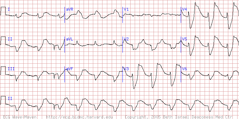 |
| Nathanson L A, McClennen S, Safran C, Goldberger AL. ECG Wave-Maven: Self-Assessment Program for Students and Clinicians. http://ecg.bidmc.harvard.edu. |
Monday, December 20, 2010
A quick look at Pulmonary Embolisms
Acute pulmonary embolism (PE) is believed to affect anywhere from 1 in 250 to 1 in 1000 persons in the US each year. Potentially 1 in 10 patients with an acute pulmonary embolism may go into cardiac arrest within the first 60 minutes[1].
The working diagnosis of a PE in the field is likely to be based solely on clinical findings. Therefore, prehospital providers should be familiar with the most common physical findings:
A combination of any of these physical and electrocardiographic findings strongly favor PE and prehospital providers should act accordingly. Unrecognized pulmonary embolisms may be rapidly fatal.
References
The working diagnosis of a PE in the field is likely to be based solely on clinical findings. Therefore, prehospital providers should be familiar with the most common physical findings:
- Tachycardia
- Tachypnea
- Dyspnea
- Persistently low SaO2
- Recent history of syncope
- Hypotension
- Cyanosis or pallor
- Diaphoresis
- Hemoptysis
- Low grade fever
- Diminished lung sounds
- Sinus tachycardia (73%)
- Prominent S1 (73%)
- "Clock-wise" rotation (56%)
- Negative T in 2+ precordials (50%)
- Incomplete or complete RBBB (20-68%)
- P-pulmonale (28-33%)
- Axis shift, generally RAD (23-30%)
- No significant findings (20-24%)
- S1Q3T3 (12-25%)
- Supraventricular arrhythmias (12%)
A combination of any of these physical and electrocardiographic findings strongly favor PE and prehospital providers should act accordingly. Unrecognized pulmonary embolisms may be rapidly fatal.
References
- Galvagno SM. Emergency Pathophysiology: Clinical Applications for Prehospital Care. Teton New Media (2003). [ISBN 1591610079]
- Surawics B, Knilans TK, Chou TC. Chou's Electrocardiography in Clinical Practice: Adult and Pediatric. Saunders/Elsevier (2008), 6th ed. [ISBN 1416037748]
Friday, December 3, 2010
Pediatric Intranasal Fentanyl
Scenario
It's a summer afternoon and you're dispatched to a 9 year old male patient involved in an ATV accident. The nearest ALS engine company has been dispatched as well. Upon your arrival you find an ATV on its side, another ATV upright, and a crowd gathered on the porch of a nearby house. A paramedic from the engine is assessing a distraught young boy, sitting in his mother's lap, holding an obviously deformed right forearm. The officer on the engine informs you that the boy and his father were riding alongside the road, traveling at 20-30 miles per hour, when the boy lost control and was thrown from the ATV (his father insists he was wearing his helmet).
You introduce yourself to the child, assuring him you're here to help, and ask him what happened. The boy states that when he fell he put his arms out and he heard a loud pop when his right hand hit the ground. He denies passing out or any other injuries but says his arm, "really hurts". He reluctantly allows you to assess his radial pulse in the affected arm, which is rapid but easily palpable. There appears to be distal involvement of both the radius and ulna, however he does not tolerate any further assessment of the arm and screams if there is any movement. The remainder of your physical exam reveals only minor abrasions to exposed skin. The engine company reports tachypnea, tachycardia, and a normal blood pressure.
Discussion
It appears the child has suffered a Colles' Fracture of the right distal forearm. Appropriate treatment would include splinting, ice packs, and pharmacologic pain control. However, given the current state of the patient, it may not be possible to splint the extremity due to anxiety and pain. Traditional prehospital pain management would require intravenous access or intramuscular administration. Both of these routes are likely to cause increased anxiety in this patient, which is best avoided.
Pain management in the pre-hospital setting is fraught with problems. Most studies have found poor provider perception of pain, underutilization of analgesics, and a hesitance to treat pediatric pain (Thomas; Greenwald). Often times, studies find that even if patients are provided analgesia, they do not feel their pain was managed adequately at all (Thomas). For pediatric patients, this problem is compounded as pre-hospital providers are often wary to provide pain management or may be unable to obtain invasive IV access to provide pain management (Greenwald). Moreover, pre-hospital providers are often placed in situations where access to patients is limited to provide pain-management, often times resulting in painful patient movements.
The addition of a noninvasive means of pain management would be an invaluable aid to pre-hospital providers and would remove a potential barrier to care. In pediatric populations, the importance of noninvasive pain management procedures is easy to grasp, as this patient population is often unable to comprehend the benefits of initially painful procedures. Improvements in "time to analgesia" will likely lead to and have a direct, positive impact on patient care and satisfaction.
Efficacy and Safety of Intranasal Fentanyl
The efficacy and safety of intranasal fentanyl (INF) has been the focus of multiple studies, both in-hospital and pre-hospital. Finn et al conducted an in-hospital randomized double-blind placebo controlled trial and found INF to have the same efficacy as oral morphine during procedural wound care in adult burn patients (n=26, 35.5 ± 12.4 years). The concentration of INF used in this study was 50 µg/mL, initial dosages of 1.48 ± 0.57 µg/kg, and no difference in the number of adverse events. Finn et al concluded that while patients receiving INF were more satisfied with their level of pain relief (p = 0.009) that overall only half of the patients in the trial reported they were "satisfied" or "very satisfied".
In a randomized, controlled, open-label study of pre-hospital INF versus IV morphine, Rickard et al found no significant difference in efficacy or safety (n=258, 42.3 ± 13.7 years). This study differs from Finn et al in that there were a multitude of chief complaints treated due to an "all-comers" design. Moreover, the doses used of INF was significantly higher at 180 µg divided evenly between the nares with up to two repeat dosages of 60 µg. Patients in the INF group received pain medication earlier than in the IV morphine group, likely owing to the simpler route of administration. Adverse effects were noted to occur more frequently in the INF group (relative risk 2.09, 95% CI 0.92-4.78, p = 0.07), however, the Rickard et al was not powered to adequately detect any statistical difference. One incidence of a significant adverse effect required a termination of the INF protocol, but it was unclear from the study if this was related to the treatment or the patient's condition. Rickard et al concluded that given the safety and efficacy of INF, it is a valuable option in patients where intravenous access is "undesirable or impossible".
Borland et al 2005 and Borland et al 2007 were inpatient randomized double-blind crossover studies evaluating the efficacy and safety of INF versus oral or IV morphine, respectively, in pediatric patients. Borland et al 2005 studied INF in pediatric burn patients requiring daily dressing changes and found no significant difference in outcomes (n=24, median 4.5 IQR 1.8-9.0 years). The INF dosage was calculated against the bioavailability of the IN route (listed as 70%) with 1.4 µg/kg fentanyl equating to an IV dosage of 1 µg/kg. There were no incidents of significant adverse events, although this was likely due to the study size. However, sedation scores recorded found that INF patients recovered earlier than their oral morphine counterparts. Overall, Borland et al 2005 found INF to be safe and efficacious, but more importantly well tolerated by pediatric patients.
Borland et al 2007 found INF to be comparable to intravenous morphine in pediatric patients presenting to the emergency department with acute long-bone fractures (n=67, 10.9 ± 2.4 years). The median total dose was 1.7 µg/kg fentanyl with repeat doses given PRN. The impetus of the study was to find alternative methods of analgesia to intravenous narcotics in the pediatric population. The study authors note that given the comparable efficacy, INF is invaluable as a means to decrease "time to analgesia" in the pediatric population with potential for pre-hospital adoption.
Mudd conducted a systematic review of the available literature for INF in the pediatric population and graded 12 studies with evidence qualities of four Level I/A, one II/A, two II/B, one III/A, and four at III/B. There was a wide variation in dosing of INF amongst the studies, with a common range of 1-2 µg/kg fentanyl. Differences in concentrations existed as well, owing to the fact that in the US fentanyl is commonly available at 50 µg/mL and is used IV/IM/IO/IN yet overseas it is often given IN with a more concentrated 100-150 µg/mL solution. No differences in significance in pain reduction were found between concentrations, only in the volume of medication delivered. While no studies found a significant difference in adverse effects, many studies had small sample sizes and no long-term studies have been completed on the action of fentanyl on the nasal mucosa. However, the evidence in the reviewed studies demonstrated three clear points: (1) that INF is as efficacious as IV/IM/PO morphine or IV fentanyl, (2) it has no difference in adverse effects, and (3) it decreases the time to analgesia administration and pain relief.
Intranasal Fentanyl Protocol
Based on the research available and the existing 2009 NC EMS protocols, an appropriate pain management protocol for the administration of intranasal fentanyl is given below:
- Adult patients with indications for narcotic analgesia for whom intravenous access is not feasible, not available, or at the discretion of the lead Paramedic, an initial dose of 50-75 µg fentanyl may be delivered intranasally. The total volume to be administered should be divided equally between the two nares (not to exceed 1mL per nare).
- If intravenous access is not available, repeat with 25 µg fentanyl delivered intranasally every 20 minutes to a maximum total dose of 200 µg.
- Pediatric patients with indications for narcotic analgesia an initial dose of 1-2 µg/kg fentanyl up to a total dose of 50 µg may be delivered intranasally. The total volume to be administered should be divided equally between the two nares (not to exceed 0.5mL per nare).
- In order to decrease the anxiety of pediatric patients requiring analgesia and invasive procedures (such as intravenous access), it may be prudent to begin with intranasal fentanyl.
- M. Borland, I. Jacobs and I. Rogers, Options in prehospital analgesia, Emerg Med (Freemantle) 14 (2002), pp. 77–84.
- M. Borland, I. Jacobs and G. Geelhoed, Intranasal fentanyl reduces acute pain in children in the emergency department: a safety and efficacy study, Emerg Med (Freemantle) 14 (2002), pp. 275–280.
- J. Finn, J. Wright, J. Fong, E. Mackenzie, F. Wood, G. Leslie and A. Gelavis, A randomized crossover trial of patient controlled intranasal fentanyl and oral morphine for procedural wound care in adult patients with burns, Burns 30 (3) (2004), pp. 262–268.
- M. Borland, R. Bergesio and E.M. Pascoe et al., Intranasal fentanyl is an equivalent analgesic to oral morphine in paediatric burns patients for dressing changes: a randomised double blind crossover study, Burns 31 (2005), pp. 831–837.
- M. Borland, I. Jacob and B. King et al., A randomized controlled trial comparing intranasal fentanyl to intravenous morphine for managing acute pain in the emergency department, Ann Emerg Med 49 (2007), pp. 335–340.
- C. Rickard, P. O’Meara, M. McGrail, et al., A randomized controlled trial of intranasal fentanyl vs intravenous morphine for analgesia in the prehospital setting, Amer J Emerg Med 25 (2007), pp. 911-917.
- S. Thomas, S. Shewakramani, Prehospital Trauma Analgesia, J Emerg Med 35 (2007), pp. 47-57.
- M. Greenwald, Analgesia for the Pediatric Trauma Patient: Primum Non Nocere? Clin Pedi Emerg Med 11 (2010), pp. 28-40.
- S. Mudd, Intranasal Fentanyl for Pain Management in Pediatrics: A Review of the Literature, J Pedi Health Care (2010), Article in Press. doi:10.1016/j.pedhc.2010.04.011.
Friday, October 22, 2010
One Year: Thank You
One year has passed since I received my EMT-Paramedic, and I'd like to say thank you.
Firstly, to my friends and family. You have endured my absence well, or at least have hid your anger well. I'm sure this last year has been tough, but probably not as tough as paramedic school. I really could not do this job without your support, especially as a volunteer. I cannot say it enough, thank you.
To my colleagues and peers, you have surely challenged me to accomplish things I never knew I was capable of doing. You have mentored me, scolded me, and sat patiently while I fumbled with IVs. There is an entire network of you online which have been invaluable as a sounding board and a reference. I can only hope I will continue to take what you have given me and make myself a better Paramedic going forward. The fact that I feel like my feet are underneath me at all is a testament to you all, thank you.
Lastly, to my patients of whom I've met quite a few: you have taught me more than I could ever hope to tell you. Some of you were thrust into my arms, others I knelt and said goodbye. You have challenged me to better myself and I appreciate every experience. My life as a green Paramedic has been an odd mix of on-the-job training for emergencies I was never told about and connecting the dots for those I was told every day about. I thank you for your understanding. I hope that I can tell a story of that time I sat next to you on a flight, and heard about your trip to see your niece get married. That is why I am here, you are why I am here. I feel blessed to meet each and every one of you, thank you.
Subscribe to:
Posts (Atom)